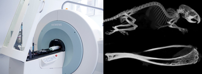
The combination of these two different imaging modalities provides a very revealing insight into the body of animals and humans.
When a plurality of CT X-rays is used to digitally visualize images, it seems that as if the body has been cut very precisely. With such cross-sectional images of all parts of the body they are represented in detail, thighs as well as skull. Unfortunately, the CT does not provide functional information.
Using a gamma camera, SPECT visualizes the low radiation from radioactive tracers inside the body. In this way it provides information about the metabolism and therefore provides information about functional processes in vivo.

Combining the two modalities has great advantages: The CT, based on imaging with X-rays is able to visualize the anatomy of the organ whereas SPECT shows the currently running processes. Both systems, X-ray unit and gamma camera are combined in one unit, their image data are combined in the common computer system. It produces very plastic 3D images that offer a wide range of information.
In clinical routine as well as basic research, this is of great use, for example during a planned lymph node surgery: The position, size and potential outbreaks can be represented as accurately before surgery that you can virtually rely on a 3D navigation device in the subsequent operation.
Furthermore the SPECT/CT system provides researchers with the ability to not only image diseases, but also to leverage the high resolution to see the unseen for more confident interpretations. Its unique quantitative capabilities provide the ability to monitor and adjust treatments earlier by accurately measuring even small differences.
